




The Disease
What is a congenital anomaly?
Congenital anomalies are structural defects affecting one or more organs. They result either from an abnormality that disrupted the normal developmental process of the embryo or fetus, or from a genetic cause. Genetic information, contained in the nucleus of every cell in the human body, is organized into 23 pairs of chromosomes, which are themselves composed of DNA and carry genes. Genetic abnormalities include:
aneuploidies (abnormalities in the number of chromosomes),
chromosomal micro-rearrangements (microdeletions, microduplications, translocation imbalances, etc.),
monogenic syndromes, whose clinical presentation may include congenital anomalies.
However, certain non-malformative genetic syndromes (such as cystic fibrosis, hemophilias, fragile X syndrome, myopathies, or metabolic syndromes) or chromosomal micro-rearrangements associated with isolated neurodevelopmental disorders are not recorded by congenital anomaly registries and, consequently, are not subject to epidemiological surveillance.
These anomalies occur primarily during the first trimester of pregnancy (the embryogenesis period) and are heterogeneous, varying in severity from minor defects to major anomalies incompatible with life.
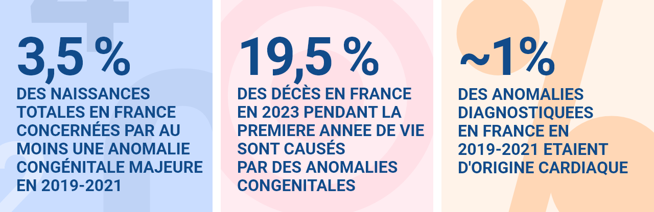
Key Statistics on Congenital Anomalies and Malformations
What are the different types of congenital anomalies?
Congenital anomalies are classified by the European EUROCAT network into groups based on common etiological mechanisms or shared clinical characteristics. They include, in particular:
Central nervous system anomalies;
ocular anomalies;
anomalies of the ears, face, and neck;
congenital heart defects;
respiratory anomalies;
oral and facial clefts;
gastrointestinal anomalies;
abnormalities of abdominal wall closure;
urinary and renal anomalies;
genital anomalies;
limb anomalies;
other anomalies/syndromes;
genetic abnormalities.
The prevalence of these anomalies varies among different groups. The most commonly diagnosed are congenital heart defects, genetic anomalies, limb anomalies, and anomalies of the kidneys and urinary tract.
The Burden of Congenital Anomalies
Congenital anomalies represent a major public health challenge: approximately 3% of births in Europe are affected. Their impact is significant, with lasting consequences for the health and quality of life of children and their families. In terms of morbidity and mortality, in France in 2023, they accounted for 24% of deaths among children under 5 and 19.5% of deaths during the first year of life (CépiDC, 2023). They also accounted for 0.68% of DALYs (disability-adjusted life years), across all ages and genders, and nearly one-fifth of these DALYs among children under 5, making them the second leading cause of DALYs in this age group (GBD, 2023).
How are they screened for and diagnosed?
Prenatal screening and diagnosis of congenital anomalies rely on medical methods that can detect, before birth, a possible morphological malformation or a genetic or chromosomal disorder. Although complementary, these two concepts must be clearly distinguished:
- The goal of prenatal screening is to estimate the risk that the fetus has an abnormality, such as trisomy 21. Certain tests are routinely offered to pregnant women.
- Prenatal diagnosis aims to confirm or rule out the presence of a suspected abnormality and to assess its prognosis. It is offered when screening reveals an increased risk or in the event of an ultrasound finding that raises concern.
Prenatal screening
Combined screening, conducted in accordance with public health policy, uses techniques that pose no risk to pregnancy. It is offered to all pregnant women, regardless of their age, and must be preceded by clear information, followed by their written consent (Article R. 2131-2 of the Public Health Code).
Since 2009, in accordance with the recommendations of the French National Authority for Health (HAS), this strategy has combined, starting in the first trimester (between 11 and 13 weeks of amenorrhea):
- an ultrasound (routine or screening) measuring nuchal translucency, to screen for certain major birth defects and to aid in the screening for chromosomal abnormalities;
- a blood test for serum marker analysis, which measures substances in the mother’s blood to assess the risk of trisomy 21 or neural tube defects (anencephaly, spina bifida).
This screening is based on a risk calculation that combines the nuchal translucency measurement, serum marker levels, and maternal age. If the calculated risk exceeds an established threshold, a confirmatory diagnostic test is recommended.
If it is not possible to perform the combined screening during the first trimester, screening can be performed later by combining:
- first-trimester ultrasound measurements and second-trimester serum marker testing;
- or second-trimester screening using serum markers alone.
DNA tests for fetal DNA circulating in the mother’s blood are highly sensitive, noninvasive tests that improve the detection rate of chromosomal abnormalities (trisomies 21, 18, and 13) and reduce the need for invasive procedures.
The HAS recommends offering these tests following a combined screening test when the estimated risk is between 1 in 1,000 and 1 in 51. However, they are not yet covered by health insurance.
- If the combined screening indicates a risk of fetal chromosomal abnormalities between 1 in 50 and 1 in 1,000, a new blood test for non-invasive prenatal testing (NIPT) is offered, followed by diagnostic confirmation via fetal karyotyping using amniotic fluid.
- If the risk is greater than 1 in 50 or if there are ultrasound findings suggesting a problem, an invasive procedure (trophoblast biopsy) is offered immediately.
Introduced in France in 2013, screening using circulating fetal DNA was expanded to include trisomies 13 and 18, and then gradually to other chromosomal abnormalities, following informed consent from the pregnant woman. Since 2017, there has been an increase in prenatal diagnoses of trisomy 21 and a decrease in invasive procedures, thanks to the widespread adoption of non-invasive prenatal testing (source: 2021 Annual Report on Prenatal Diagnostic Activities, Biomedicine Agency).
In addition to screening, at least two ultrasounds are recommended in France:
- The second-trimester morphological ultrasound (around 22 weeks): This helps detect malformations that were not previously identified. Its findings may lead to consideration of pregnancy termination, specific treatment after birth, or in utero treatment.
- The third-trimester ultrasound: This evaluates fetal growth and detects any late-onset abnormalities.
Prenatal Diagnosis
Prenatal diagnosis is offered to pregnant women at increased risk of fetal abnormalities, identified either:
- before pregnancy (following a genetic counseling consultation, in cases of a family history or risk of transmission of a genetic abnormality);
- during pregnancy (following an abnormal ultrasound finding or an elevated risk identified during organized screening, such as for Down syndrome).
It relies on additional tests (imaging or laboratory tests, such as a blood test or amniocentesis) and aims to:
- confirm or rule out the suspected abnormality;
- assess the prognosis;
- adjust prenatal care and plan for the care of the unborn child;
- help parents prepare for the child’s arrival.
A clinical genetics consultation may be offered to a pregnant woman, following her referral by a CPDPN, when a screening test indicates a suspected abnormality in the unborn child. It is also offered to couples for whom a risk was identified prior to pregnancy, based on the parents’ medical history.
These consultations are conducted by a medical geneticist or a genetic counselor (under the supervision of the geneticist). They allow for:
- to guide any genetic testing of the fetus (cytogenetic or molecular genetic analyses);
- to determine the severity of suspected or diagnosed conditions;
- to inform the parents about treatment options or, if applicable, the possibility of a medical termination of pregnancy, if that is their wish.
The diagnosis of a genetic abnormality is based on various tests:
- Cytogenetics: karyotyping, DNA microarray analysis (ACPA), FISH.
- Molecular genetics: targeted analysis of a single gene or a panel of genes, or pan-genomic analyses (exome or genome sequencing).
These tests may be offered prenatally or postnatally, depending on the clinical context and the parents’ wishes. The procedures for accessing prenatal diagnosis are defined in the Public Health Code (Articles R2131-1 through R2131-9-1).
Postnatally, a genetic diagnosis can be made remotely, particularly thanks to easier access to genome sequencing since 2020, as part of the France Genomic Medicine Plan 2025 (for indications selected by the HAS). Parents remain free to decline this offer. It is also possible to update the registry databases once the results are known, with no time limit after inclusion.
When the estimated risk is greater than or equal to 1 in 250, diagnostic confirmation is offered via fetal karyotyping, which requires an invasive procedure:
- Chorionic villus sampling (collection of cells from the developing placenta): can be performed as early as the 12th week of amenorrhea.
- Amniocentesis (collection of amniotic fluid): can be performed starting at the 16th week of amenorrhea.
- Cordocentesis (collection of fetal blood): can be performed after the 20th week of amenorrhea, under very specific indications.
These procedures carry a risk of miscarriage (0.5% for amniocentesis, 1% for trophoblast biopsy, and higher for cordocentesis).
In France, the CPDPNs bring together experts in fetal medicine (obstetrician-gynecologists, geneticists, ultrasound specialists, and pediatricians/neonatologists). Their mission is to:
- to certify, following a multidisciplinary evaluation, that a fetal anomaly presents a “high probability that the unborn child will suffer from a particularly serious condition deemed incurable at the time of diagnosis ” (Art. L. 2231-1 of the CSP):
- to allow, if the woman or the couple so desires, a medically indicated termination of pregnancy;
- to contribute to the monitoring of the pregnancy, the delivery, and the care of the newborn under the best possible conditions.
Learn more about multidisciplinary prenatal diagnosis centers | Agence de la Biomédecine
In relation to

Tobacco
thematic dossier
Despite a slight decrease compared to 2015, tobacco remains responsible for more than 68,000 premature deaths in 2023—accounting for 11% of total mortality—making it still the leading cause of...

Alcohol
thematic dossier
Alcohol is deeply rooted in French culture. Its consumption remains a major cause of death and illness in France.

Pesticides
thematic dossier
Pesticides are widely used in France, primarily in agriculture, and the population is heavily exposed to them. Their effects on the health of the general population are still poorly understood.